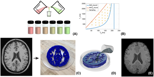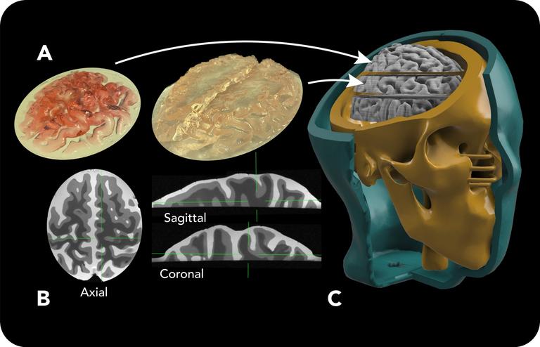
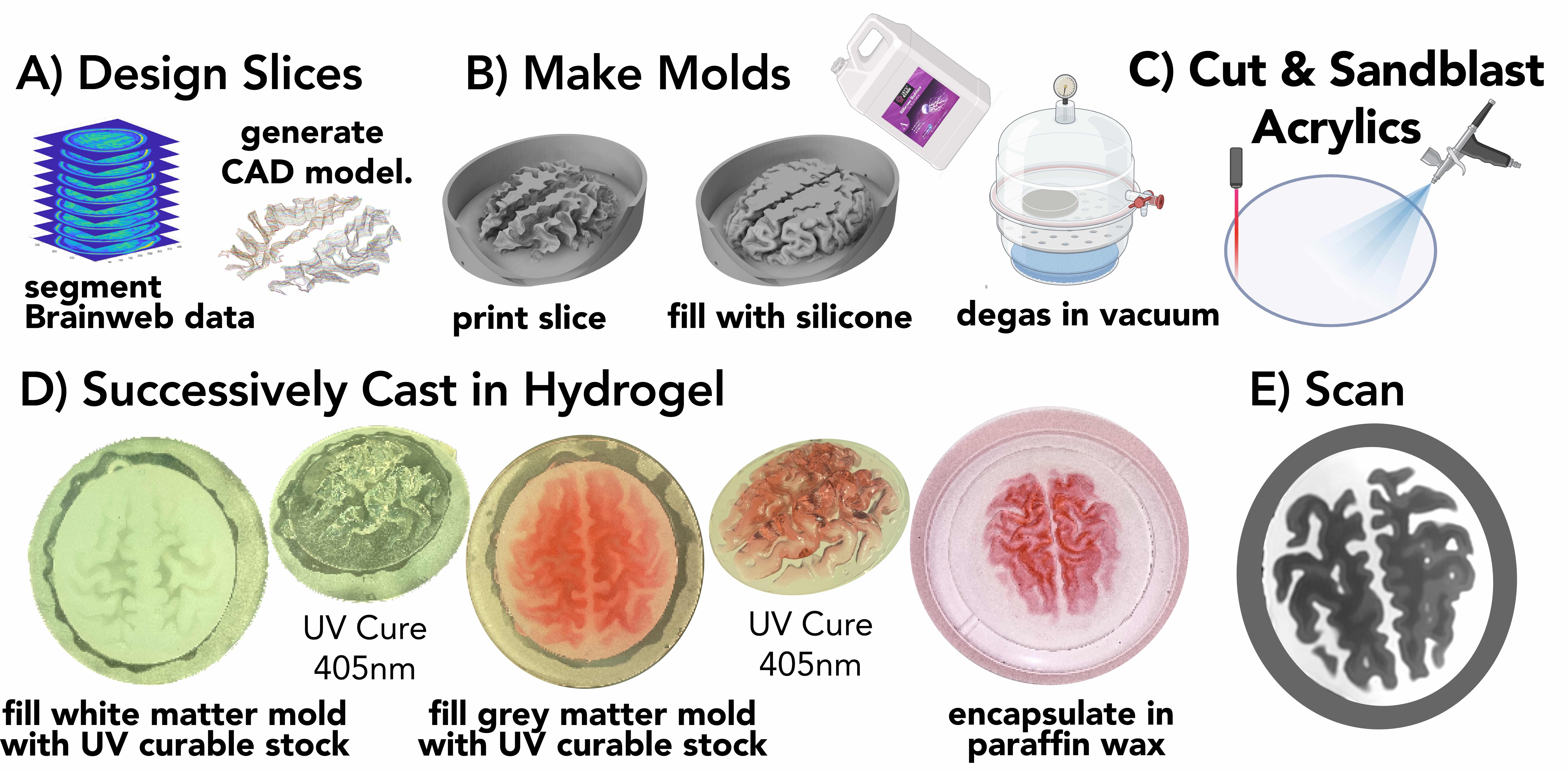
Magnetic resonance imaging (MRI) is a safe and powerful imaging modality. Researchers are constantly developing new methods and techniques to make imaging faster and higher quality. To validate these methods they need phantoms; devices used in place of a live subject. As MRI, specifically quantitative MRI, evolves and grows, so does the need for reliable and realistic quantitative phantoms. Generally, MRI phantoms consist of fluid or gel containing ions or nanoparticles, sealed into containers with simple geometry, such as cylinders or spheres. Current research focuses on developing standardized, quantitative phantoms that are biologically accurate. The sophistication of these phantoms is generally limited because it is difficult to achieve anatomical 3D geometry and mimic the contrast of multiple tissue types, especially without boundaries between sections. Our project focuses on achieving highly realistic anatomical geometry in combination with biological contrast values to create phantoms that are precise and quantitative. These highly precise phantoms present potential for accelerated development of quantitative MRI, as they are used for machine calibration, technological development, and can be used in the place of human test subjects.
Key Contributions
- Realistic Anatomical Geometry: Developed phantoms with highly realistic 3D anatomical structures that closely mimic human anatomy for more accurate MRI validation
- Biological Contrast Values: Created phantoms with biologically accurate contrast values across multiple tissue types without artificial boundaries between sections
- Quantitative Accuracy: Achieved precise calibration capabilities enabling more reliable quantitative MRI measurements and method validation
- Advanced Manufacturing: Integrated sophisticated fabrication techniques to create complex anatomical geometries while maintaining biological accuracy
People

Izabel Wu
StaffRelated Publications
Last updated: 2025-09-03

